A prognosis of blindness comes as a hard blow. It signals a future of extreme challenges and presupposes extraordinary effort required to maintain one's independence by adapting to dramatically changed circumstances. For those diagnosed with diseases such as age-related macular degeneration (AMD) or retinitis pigmentosa (RP), both of which affect the retina, the light-sensitive layer of tissue at the back of the eye, that prognosis is all too common. AMD is prevalent in people over 50, and RP can strike at virtually any stage in life.
At UC Santa Barbara, decades of research into the cell biology of the eye have led to potential therapies that are currently undergoing clinical trials to determine whether they are safe and effective enough to be used to treat patients. If they meet stringent FDA standards, the proposed therapies — developed under the leadership of UCSB researchers in the Center for Stem Cell Biology and Engineering at the Neuroscience Research Institute (NRI), working in collaboration with scientists from USC, City of Hope, and UC Irvine — could be used to treat blindness in those afflicted with AMD and RP.
AMD: Blurs and Blank Spots
Those suffering from AMD may see the world as if they were looking through a camera that has a large, nearly opaque smudge on the lens or the viewfinder. The center is obscured, while the periphery remains sharp. Text may be blurry or illegible, straight lines may appear to be wavy, and colors may lose vibrancy.
“It devastates your ability to carry out everyday tasks,” says Dennis Clegg, a neurobiologist and Wilcox Family Chair in BioMedicine at UCSB. If the disease is left untreated, the dim haze in the center of the patient’s visual field eventually becomes a large blind spot, making tasks like reading, writing, getting an object from a cupboard, recognizing faces, and even walking in a familiar space nearly impossible.
Clegg explained that AMD begins with the death of a layer of cells in the back of the eye called the retinal pigmented epithelium (RPE). “The retinal pigmented epithelium is crucial to the support of photoreceptors — the light-sensitive cells that are responsible for our ability to detect light and form a visual image,” he explained. “Eventually, the photoreceptors also die.” The disease also severely affects the photoreceptors within the macula, a region of cells less than one millimeter in diameter located near the center of the retina and responsible for high-acuity vision.
“ What’s most interesting about the retina is that when you look at it, you're actually seeing a part of the brain.”
The cause of AMD is not entirely clear, Clegg says. But recent research, including that conducted by UCSB emeritus research professors Lincoln Johnson and Don Anderson, recast AMD as an inflammatory disease that affects the complement system — a component of the body’s immune system — likely leading to dysfunction and degeneration of RPE cells. Further research has identified variants in complement-system genes that are specifically linked to increased risk for AMD.
Whatever the key factor, AMD has become one of the leading causes of progressive, irreversible loss of vision in millions of elderly Americans. The National Eye Institute projects that by 2030, nearly four million Americans over the age of fifty will have AMD.
Seeking the Cause — TESTING a Cure
The quest for a cause of, and a potential cure for, AMD began in earnest at UCSB about twenty years ago, when the Center for the Study of Macular Degeneration (CSMD) was established within NRI. In a joint research effort, Johnson, Anderson, and collaborator Gregory Hageman — now a professor of ophthalmology and visual sciences at the University of Utah — laid out the potential role of immune reaction and inflammation as causes of the RPE cell death.
At around the same time, interest in stem cell research had taken hold. Scien-tists realized the vast potential of the cells, including their ability to repair damaged organs, especially those in the nervous system, which has a limited capacity for self-repair.
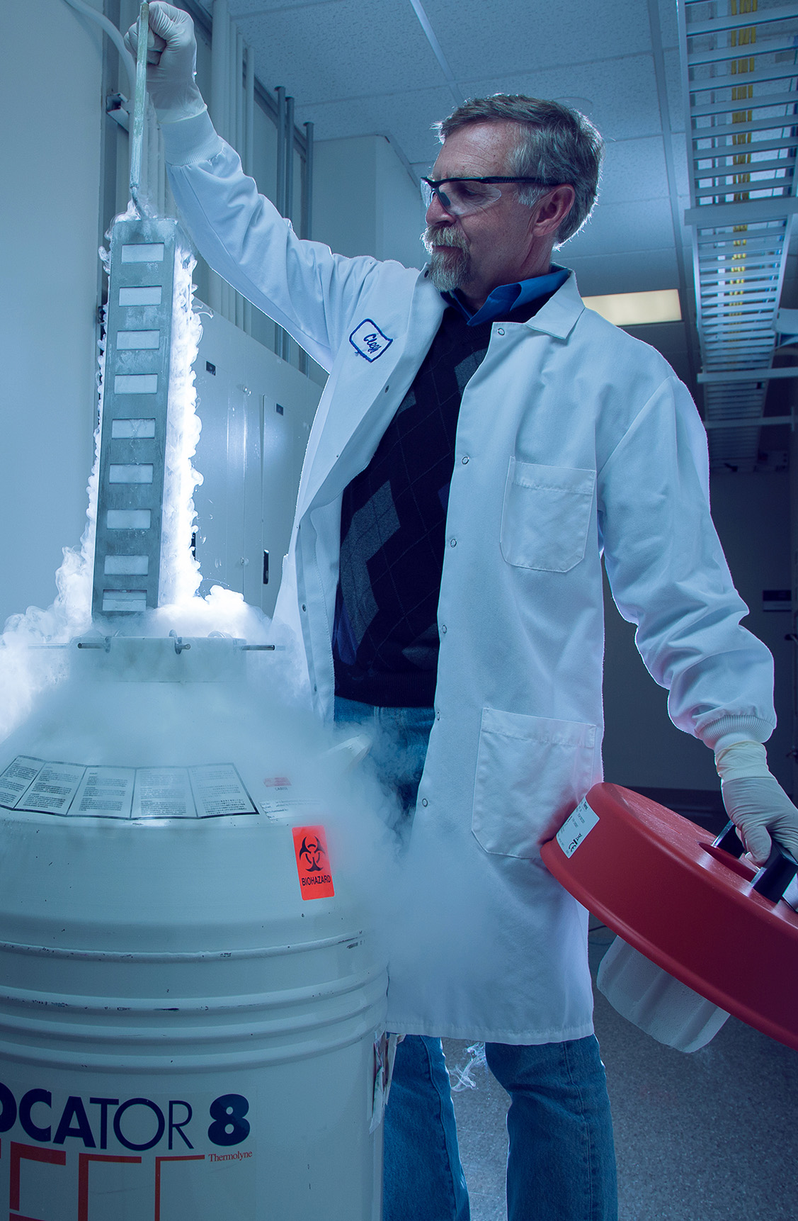
 Professor Dennis Clegg retrieves frozen stem cells from a liquid-nitrogen storage tank in UCSB's Center for Stem Cell Biology and Engineering.
Clegg was also interested in the neural aspect of AMD. “For me, the whole reason for conducting basic research to study neural development on a molecular and cellular basis was that it might someday be relevant in treating a disease,” he said.
In 2004, California voters approved an initiative that led to the creation of the California Institute for Regenerative Medicine (CIRM), an agency to fund stem cell research. That year, Clegg said, research also showed that stem cells could be prompted to make RPE cells, the very ones destroyed by AMD.
With funding from CIRM, Clegg and Johnson initiated a collaboration with USC researchers Mark Humayun and David Hinton, Jane Lebkowski at Geron Corporation in Menlo Park, City of Hope scientists, and Caltech engineers. The team, named the California Project to Cure Blindness, developed a patch consisting of stem cell-derived RPE arrayed on a scaffold, and devised a way to insert it into the eye of an AMD patient.
The surgical procedure, which has been performed on two patients in the clinical trial, involves using a specially designed instrument to insert the patch behind the retina. The RPE cells, arranged like a layer of tiny Legos, fits right onto the photoreceptors, providing them with much-needed nutritional and physiological support. The polymer scaffold used to support the RPE cell layer is made of parylene, which is also used in pacemakers and other implants.
“The first patient, an 83-year-old woman, really wanted to be the first,” Clegg said, adding, “Those who volunteer for a clinical trial are my heroes.” The group hopes to perform the procedure a few more times before reporting their progress. Once the procedure has been proven safe, the next phase will be to test it on patients who are in earlier stages of the disease.
RP: Night Blindness and Tunnel Vision
To Steven Fisher and Geoff Lewis — scientists studying retinal cell biology in UCSB’s Neuroscience Research Institute — what’s most interesting about the retina is that when you look at it, you’re actually seeing a part of the brain.
“It’s a beautiful structure,” Lewis said, who, like Fisher, has been studying the retina for more than thirty years. “It’s somewhat simplified compared to the brain in that it has only two synaptic layers and three cell-body layers, but it’s also an accessible part of the central nervous system that allows for easier manipulation compared to the brain or the spinal cord.”
The retina can also degenerate as a result of a host of genetic conditions that fall under the umbrella of retinitis pigmentosa (RP).
Fisher explained RP as a family of diseases that involve over a hundred different mutations and a variety of molecules, all of which give rise to similar symptoms. While the timing, effects, and severity may vary, he said, the sequence of photoreceptor degeneration is the same. With RP, the rod photoreceptors — responsible for vision in low-light conditions and the detection of motion — die first, causing night blindness and tunnel vision. The disease eventually causes the color-sensitive cones to die as well. According to the National Eye Institute, RP, which is both genetically inherited and incurable affects an estimated 1 in 4,000 people worldwide. Fisher and Lewis want to help them by finding ways to rescue those dying cells.
“If you can slow or prevent the degeneration of the rod photoreceptors that form peripheral vision, then you can perhaps maintain central vision,” said Lewis, who focused his past research on retinal detachment and re-attachment.
At around the same time, and from the same effort that resulted in the CIRM funding for AMD research, Lewis and Fisher encountered like-minded UC Irvine neurobiologist and ophthalmologist Henry Klassen, who had spent decades conducting studies and experiments on retinal diseases. With funding from CIRM, the three began collaborative experiments to pursue Klassen’s main idea, which was to inject retinal progenitor cells into the affected eye to determine whether they could rescue the failing photoreceptors.
Progenitor cells, Fisher explained, are stem cells that have partially differentiated so that they lose their “pluripotent” potential, but have not yet assumed the final differentiated state of adult cells.
“They are stem cells that are taken along the lineage, essentially becoming cells that will give rise to the cells, in, say, the eye. So they’re taken further along the differentiation line,” he noted.
“But we don’t necessarily want them to differ-entiate,” Lewis added. “Ideally we want them to remain undifferentiated so that they continue to secrete biochemicals that may rescue the photoreceptors.” The progenitor cells, according to the procedure developed by Klassen, are injected into the vitreous — the clear gel between the retina and the lens of the eyeball — where they secrete molecules that protect the ailing photoreceptors and prevent further degeneration.
Fisher and Lewis’s task was to assess the safety and efficacy of the procedure in animal models of RP as it made its way to clinical trials, which began in late 2015. Aside from confirming that the small cluster of about 100,000 cells would actually do its job, they also wanted to rule out potential problems, such as the formation of tumors and other undesirable effects.
Now, after a year of trials, and having treated one eye in each member of the first cohort of RP patients, all considered legally blind, the researchers have seen great progress.
“It’s so heartwarming to hear these patients talk about how they went from darkness essentially to now seeing almost too much light compared to what they were used to,” said Lewis. “Some are even wearing sunglasses now and saying that they are starting to see color again.”
Patients are reporting not only that progression toward total blindness has slowed or stopped, but also that they are seeing signs of reversal of the disease, which may represent a revival of the non-functional photoreceptors.
Questions remain regarding the new treatment, such as how long the progenitor cells will continue to be effective and whether patients would require additional injections to maintain their vision.
“We don’t know the answer to that,” Lewis said. “It’s possible that after a year or two you’ll have to re-inject additional cells, but injections into the eye are not uncommon, so we don’t anticipate any problems.”
As they embark on the next phase of clinical trials, Fisher and Lewis are also looking at whether this type of technology might apply to other diseases of the retina that result in cell death, such as glaucoma or diabetic retinopathy.“There are so many diseases that cause the cells in the eye to die and lead to blindness,” Fisher explained.
Should this therapy prove successful in treating what is a family of diseases that have genetic origins, he added, it may prove a “universal fit” for other diseases that aren’t the result of a malfunctioning gene. “The possibility is that this will carry over,” Fisher said. “That’s the big hope.”

Postdoctoral researcher Britney Pennington prepares cell culture medium to differentiate stem cells into RPE.
